Drawing Of Prophase
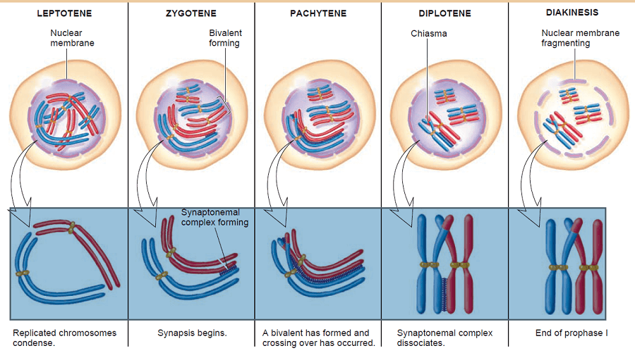
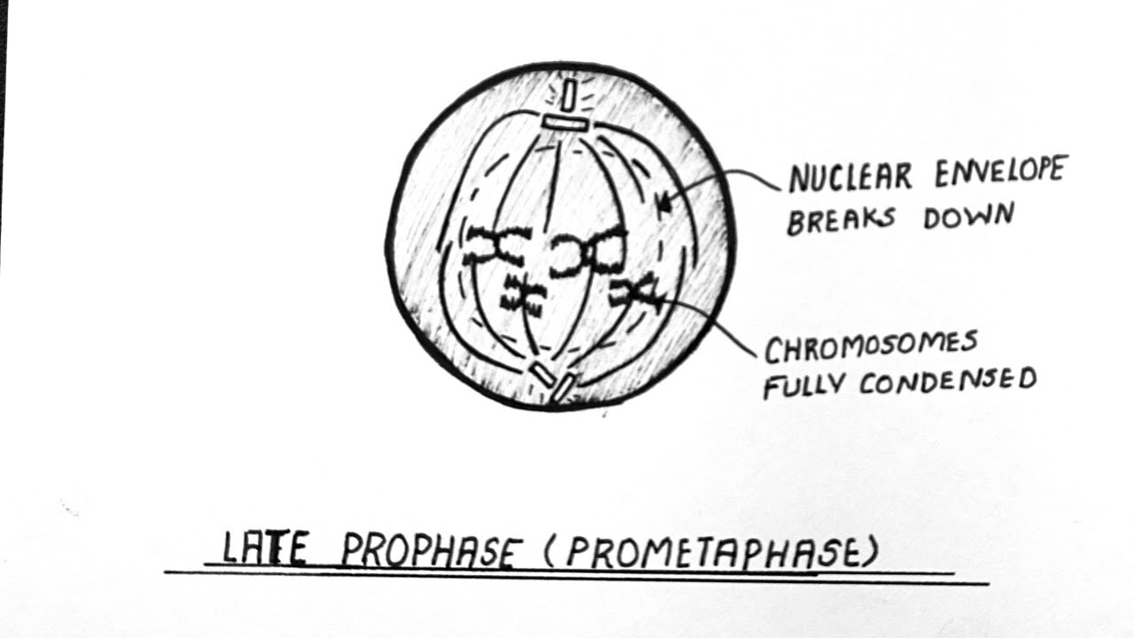
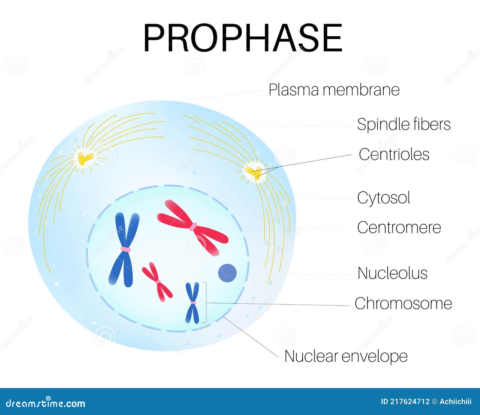
Drawing Of Prophase - Some textbooks list five, breaking prophase into an early phase (called prophase) and a late phase (called prometaphase). During prophase i, chromosomes pair up and exchange genetic material, creating more variation. During prophase, the parent cell chromosomes — which were duplicated during s phase —. Web prophase is the first phase of mitosis, the process that separates the duplicated genetic material carried in the nucleus of a parent cell into two identical daughter cells. Web prophase (versus interphase) is the first true step of the mitotic process. During prophase i, differences from. Prophase, metaphase, anaphase, and telophase. Beginning after interphase, dna has already been replicated when the cell enters prophase. Nuclear membrane breaks down, chromatin condenses, mitotic spindle forms and attaches to kinetochores. Microtubules align chromosomes along metaphase plate. Nuclear membrane breaks down, chromatin condenses, mitotic spindle forms and attaches to kinetochores. Prophase i is the first stage of meiosis i, followed by prophase ii, anaphase i, anaphase ii, metaphase i and metaphase ii. The mitotic spindle, composed of microtubules and proteins, forms in the cytoplasm. Web prophase is the first stage in mitosis, occurring after the conclusion of the g 2 portion of interphase. In metaphase i, chromosomes line up in the middle of the cell. These phases are prophase, prometaphase, metaphase, anaphase, and telophase. During prophase, the parent cell chromosomes — which were duplicated during s phase —. During interphase, the parent cell’s chromosomes are replicated, but they aren’t yet visible. Some textbooks list five, breaking prophase into an early phase (called prophase) and a late phase (called prometaphase). Mitosis, a key part of the cell cycle, involves a series of stages (prophase, metaphase, anaphase, and telophase) that facilitate cell division and genetic information transmission. In animal cells, the centrioles near the nucleus begin to separate and move to opposite poles of the cell. The mitotic spindle, composed of microtubules and proteins, forms in the cytoplasm. During prophase, several important changes occur: Web prophase is the phase that follows the interphase and typically the first and longest phase in the cell cycle, for both mitosis. Web the first and longest phase of mitosis is prophase. Prophase is the first step of mitosis. Nuclear membrane breaks down, chromatin condenses, mitotic spindle forms and attaches to kinetochores. Web prophase, in both mitosis and meiosis, is recognized by the condensing of chromosomes and separation of the centrioles in the centrosome. Web today, mitosis is understood to involve five. Web the prophase under a microscope shows the gradually becoming condensed chromatin, resulting in the formation of the individual chromosome. Web prophase (versus interphase) is the first true step of the mitotic process. In meiosis i, cells go through four phases: Centrosomes and microtubules play pivotal roles in orchestrating this complex process, ensuring the successful replication of cells. As in. Prophase is the first step of mitosis. In animal cells, the centrioles near the nucleus begin to separate and move to opposite poles of the cell. In metaphase i, chromosomes line up in the middle of the cell. Prophase, metaphase, anaphase, and telophase. Web prophase is the first step of mitosis. Some textbooks list five, breaking prophase into an early phase (called prophase) and a late phase (called prometaphase). During prophase, the parent cell chromosomes — which were duplicated during s phase —. During prophase i, chromosomes pair up and exchange genetic material, creating more variation. Microtubules align chromosomes along metaphase plate. Nuclear membrane breaks down, chromatin condenses, mitotic spindle forms. Nuclear membrane breaks down, chromatin condenses, mitotic spindle forms and attaches to kinetochores. In meiosis i, cells go through four phases: Imagine the difference between a slinky fully stretched out, and a slinky that has been pressed back together. Web prophase is the first step of mitosis. You know this prophase is the first stage of mitosis cell division which. Some textbooks list five, breaking prophase into an early phase (called prophase) and a late phase (called prometaphase). Web the prophase under a microscope shows the gradually becoming condensed chromatin, resulting in the formation of the individual chromosome. Web prophase (versus interphase) is the first true step of the mitotic process. These phases are prophase, prometaphase, metaphase, anaphase, and telophase.. Web prophase, the initial stage of mitosis and of the mitotic division of meiosis, characterized by the formation of the mitotic spindle and the condensation of the chromosomes. In metaphase i, chromosomes line up in the middle of the cell. Microtubules align chromosomes along metaphase plate. During interphase, the parent cell’s chromosomes are replicated, but they aren’t yet visible. It. In animal cells, the centrioles near the nucleus begin to separate and move to opposite poles of the cell. During prophase, several important changes occur: Centrosomes and microtubules play pivotal roles in orchestrating this complex process, ensuring the successful replication of cells. You know this prophase is the first stage of mitosis cell division which may quickly identify with the. Centrosomes start to form structures which help the cell through the rest of mitosis. Prophase is the first step of mitosis. Web prophase is the first stage in mitosis, occurring after the conclusion of the g 2 portion of interphase. Mitosis begins at prophase with the thickening and coiling of the chromosomes. In metaphase i, chromosomes line up in the. Imagine the difference between a slinky fully stretched out, and a slinky that has been pressed back together. Web prophase is the first stage in mitosis, occurring after the conclusion of the g 2 portion of interphase. Mitosis begins at prophase with the thickening and coiling of the chromosomes. It is the phase of dna unwinding and chromatin condensation to make the chromosomes visible. During interphase, the parent cell’s chromosomes are replicated, but they aren’t yet visible. During prophase i, differences from. Web prophase, the initial stage of mitosis and of the mitotic division of meiosis, characterized by the formation of the mitotic spindle and the condensation of the chromosomes. During prophase, several important changes occur: Web prophase, in both mitosis and meiosis, is recognized by the condensing of chromosomes and separation of the centrioles in the centrosome. During prophase i, chromosomes pair up and exchange genetic material, creating more variation. Web the first and longest phase of mitosis is prophase. Centrosomes start to form structures which help the cell through the rest of mitosis. These phases are prophase, prometaphase, metaphase, anaphase, and telophase. This organelle controls the microtubules in the cell, and each centriole is one half of the organelle. Web the prophase under a microscope shows the gradually becoming condensed chromatin, resulting in the formation of the individual chromosome. Some textbooks list five, breaking prophase into an early phase (called prophase) and a late phase (called prometaphase).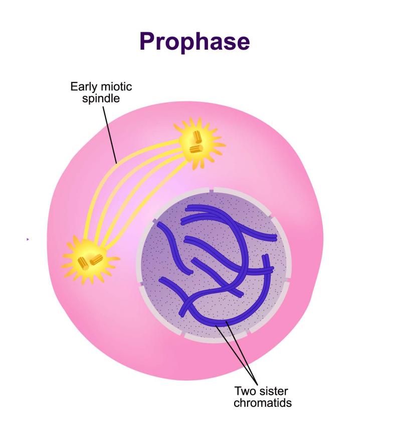
Prophase Diagram
Prophase in mitosis and meiosis (Prophase 1 and 2)

How to draw easily PROPHASE 1 OF MEIOSIS 1 / PROPHASE 1/ CELL DIVISION
Prophase Diagram How To Draw Labelled Diagram Of Prophase Class
Draw The Diagram Of All Five Sub Stages Of Prophase 1 Of
Prophase is the Phase of the Cell Cycle. Stock Vector Illustration of

Prophase Diagrams

Prophase Tutorial Sophia Learning
Prophase is the first stage of cell division. 14268877 Vector Art at
Prophase Diagrams
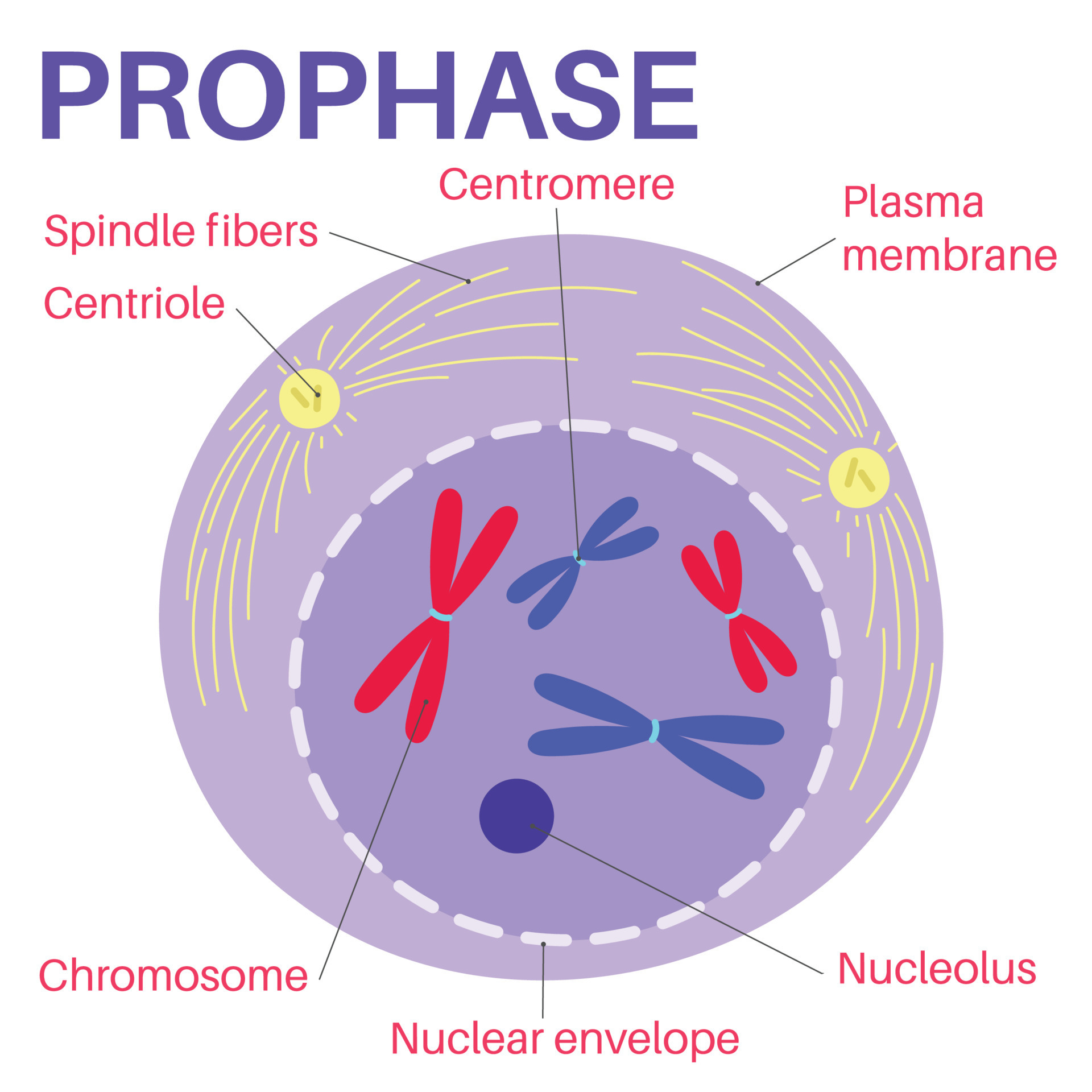
In Animal Cells, The Centrioles Near The Nucleus Begin To Separate And Move To Opposite Poles Of The Cell.
You Know This Prophase Is The First Stage Of Mitosis Cell Division Which May Quickly Identify With The Help Of A Light Microscope.
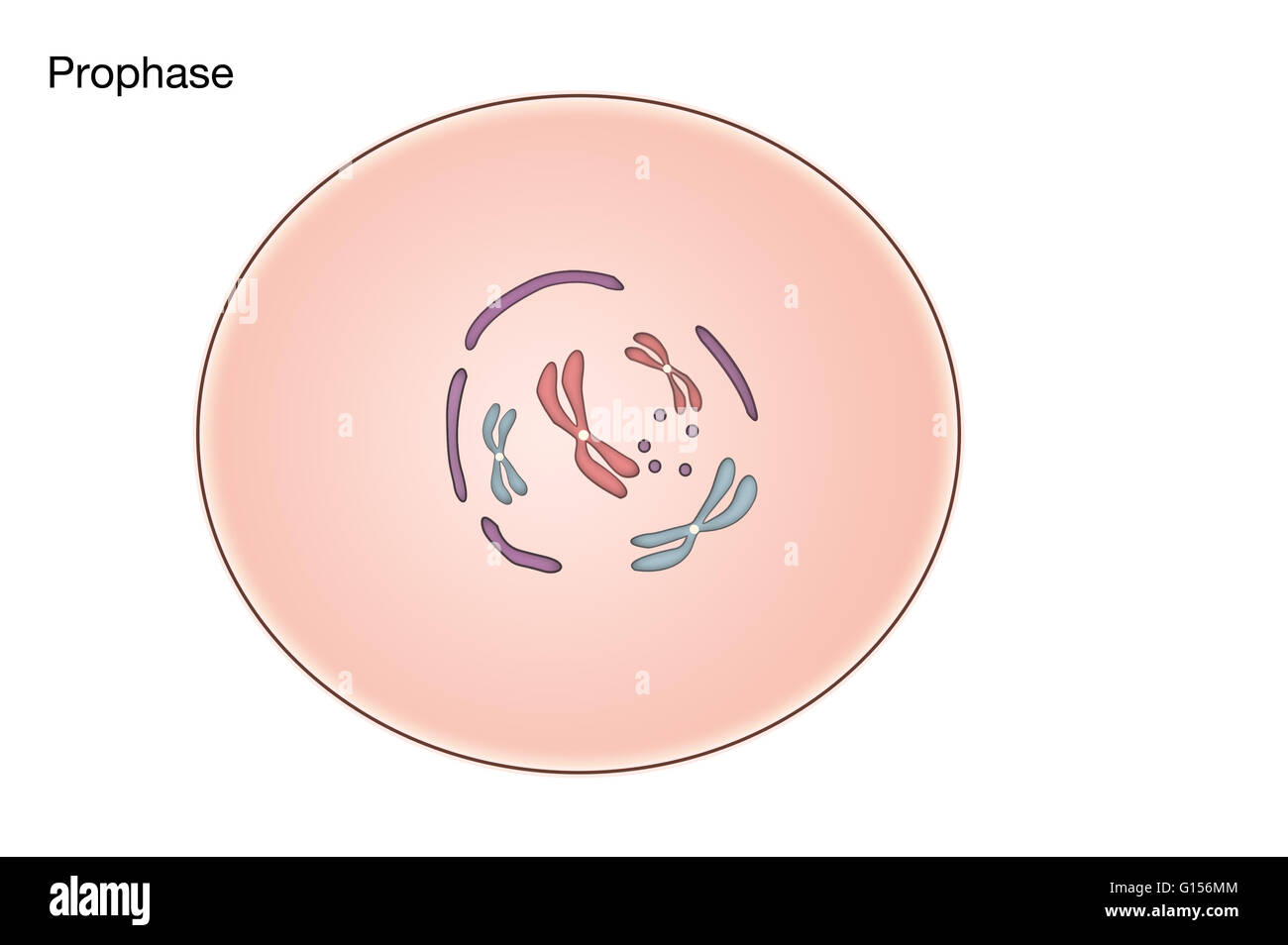
This Is When The Genetic Fibers Within The Cell’s Nucleus, Known As Chromatin, Begin To Condense And Become Tightly Compacted Together.
In Metaphase I, Chromosomes Line Up In The Middle Of The Cell.
Related Post: