Drawing Of Gross Anatomy Of The Urinary System
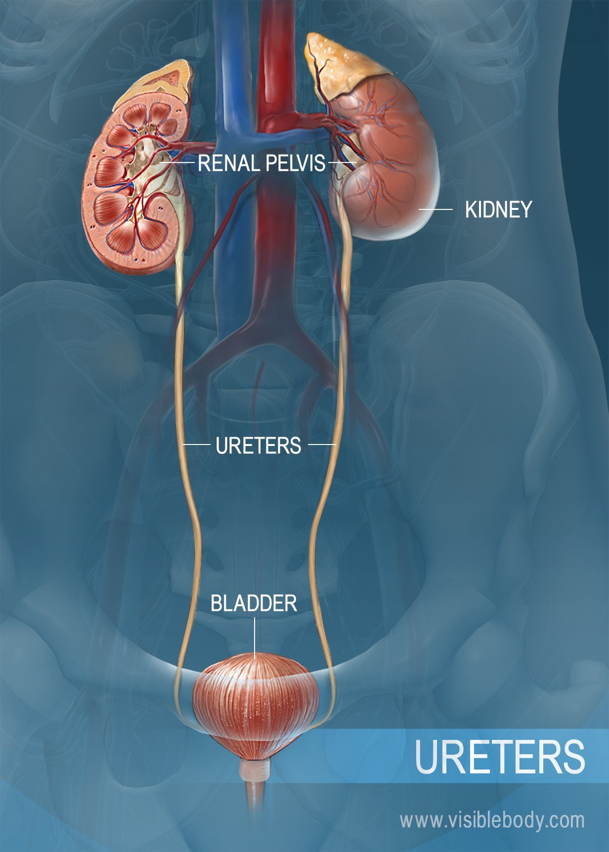
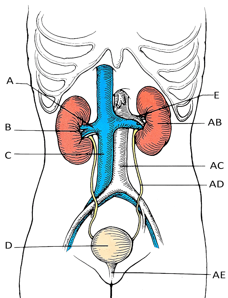
Drawing Of Gross Anatomy Of The Urinary System - Excretory products and their elimination To be able to pee, your body must pass this waste through a series of organs, ducts and tubes. Web you read about the different parts of the human urinary system with a diagram in detail. Kidneys, ureters, urinary bladder, and urethra. Supplied by renal artery and vein. Specifically, you will examine the gross and microscopic anatomy of the system as it is represented in humans. Hilum is medial cleft for vessels and ureters to pass. The renal columns are connective tissue extensions that radiate downward from the cortex through the medulla to separate the most characteristic features of the medulla, the. View detailed illustrations of the kidneys, bladder, and other urinary system structures. Web part of the teachme series. Identify the ureters, urinary bladder, and urethra, as well as their location, structure, histology, and function. By the end of this section, you will be able to: Describe voluntary and involuntary neural control of micturition. The urinary system (or urinary tract) works as your body’s filtration system. Web in this article we’ll be walking you through the best way to learn the anatomy of the urinary system: Name structures found in the cortex and medulla. The ureters are typically 28 to 34 centimeters in length and are positioned posterolateral in the body. Kidneys, ureters, urinary bladder, and urethra. Practice tests, labeling quizzes and diagrams abound! When your urinary system removes toxins and wastes from your body, it comes out as pee (urine). For related information on other topics, visit byju’s. The urinary system consists of two kidneys, two ureters, a urinary bladder, and a urethra. The body takes nutrients from food and converts them to energy. Practice tests, labeling quizzes and diagrams abound! Specifically, you will examine the gross and microscopic anatomy of the system as it is represented in humans. You will also learn how blood flow through the. Web this chapter will help you to understand the gross and microscopic anatomy of the urinary system: Describe voluntary and involuntary neural control of micturition. Web study with quizlet and memorize flashcards containing terms like renal artery, renal hilum, kindey and more. Web anatomy of the urinary system. For related information on other topics, visit byju’s. Specifically, you will examine the gross and microscopic anatomy of the system as it is represented in humans. Web anatomy of the urinary system. Web anatomy of the urinary system. Identify the major blood vessels associated with the kidney and trace the path of blood through the kidney. Web identify the major internal divisions and structures of the kidney. Web study with quizlet and memorize flashcards containing terms like renal artery, renal hilum, kindey and more. Paired, right is lower than left. Identify the ureters, urinary bladder, and urethra, as well as their location, structure, histology, and function. The kidneys alone perform the functions just described and manufacture. By the end of this section, you will be able to: Paired organs located near the lower back that are the primary sites for blood filtration and urine formation. Web key features of the urinary system: Specifically, you will examine the gross and microscopic anatomy of the system as it is represented in humans. Web anatomy of the urinary system. Identify the ureters, urinary bladder, and urethra, as well as their location, structure, histology, and function. The ureters are bilateral tubes that drains urine from the kidneys to the bladder through peristalsis and gravity (kowalczyk, 2022). Describe voluntary and involuntary neural control of micturition. The organs of the urinary system include the kidneys, renal pelvis, ureters, bladder and urethra. To. Describe voluntary and involuntary neural control of micturition. Web part of the teachme series. The medical information on this site is provided as an information resource only, and is not to be used or relied on for any diagnostic or treatment purposes. Identify the ureters, urinary bladder, and urethra, as well as their location, structure, histology, and function. Identify the. Paired organs located near the lower back that are the primary sites for blood filtration and urine formation. You will also learn how blood flow through the. The kidneys alone perform the functions just described and manufacture urine in the process, while the other organs of the urinary system provide temporary storage reservoirs for urine or serve as. It is. Compare and contrast male and female urethras. Web in this laboratory, you will use models, diagrams and histological samples to study the anatomy of the urinary system. Web anatomy of the urinary system. Test your knowledge of the urinary system with our unlabeled diagram: Web this chapter will help you to understand the gross and microscopic anatomy of the urinary. The ureters are bilateral tubes that drains urine from the kidneys to the bladder through peristalsis and gravity (kowalczyk, 2022). Identify the ureters, urinary bladder, and urethra, as well as their location, structure, histology, and function. The medical information on this site is provided as an information resource only, and is not to be used or relied on for any. Compare and contrast male and female urethras. When your urinary system removes toxins and wastes from your body, it comes out as pee (urine). Practice tests, labeling quizzes and diagrams abound! View detailed illustrations of the kidneys, bladder, and other urinary system structures. The blood to produce urine. A muscular sac that stores urine until it is expelled from the body. The body takes nutrients from food and converts them to energy. The kidneys alone perform the functions just described and manufacture urine in the process, while the other organs of the urinary system provide temporary storage reservoirs for urine or serve as. Compare and contrast the cortical and juxtamedullary nephrons. Paired organs located near the lower back that are the primary sites for blood filtration and urine formation. Web the urinary bladder is a hollow, muscular, and distensible organ that sits on the pelvic floor and below the peritoneum. Test your knowledge of the urinary system with our unlabeled diagram: Web key features of the urinary system: The urinary system consists of two kidneys, two ureters, a urinary bladder, and a urethra. The medical information on this site is provided as an information resource only, and is not to be used or relied on for any diagnostic or treatment purposes. Excretory products and their elimination
Male Urinary System Medical Stock Images Company
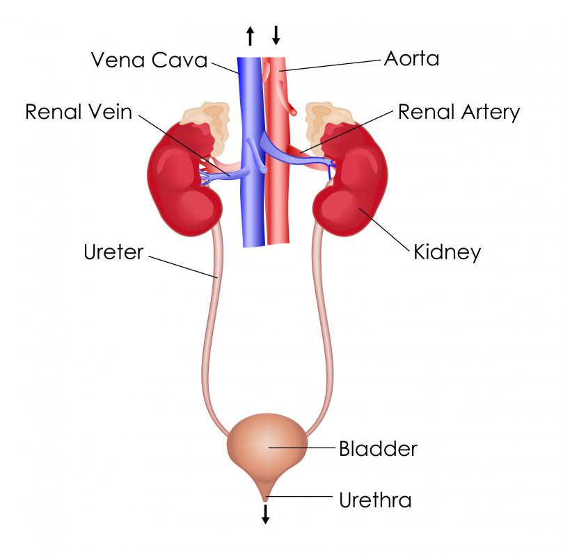
What Is the Structure of the Urinary System? (with pictures)
Urinary System Structures
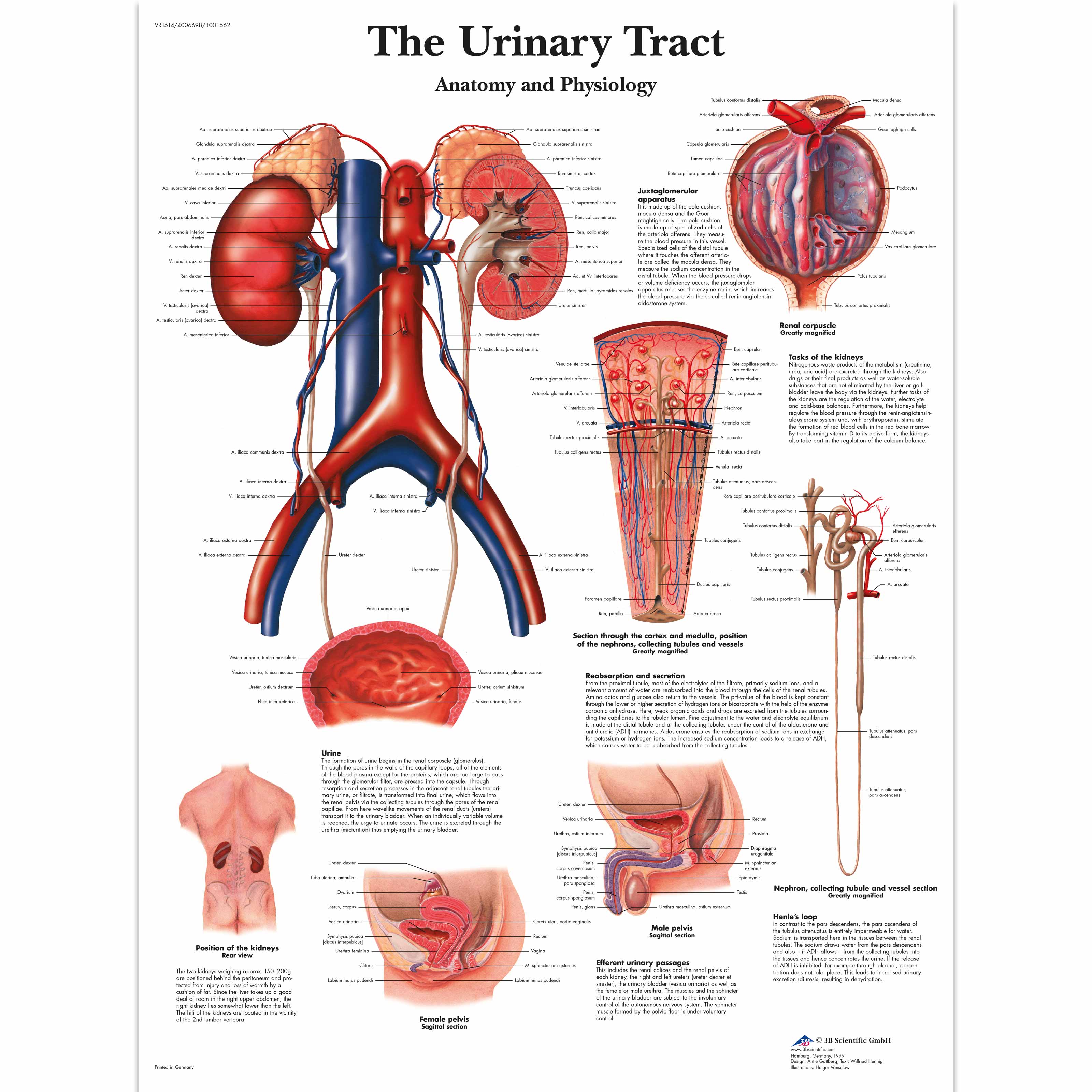
The Urinary Tract Anatomy and Physiology 1001562 3B Scientific
Urinary System Diagram World of Reference
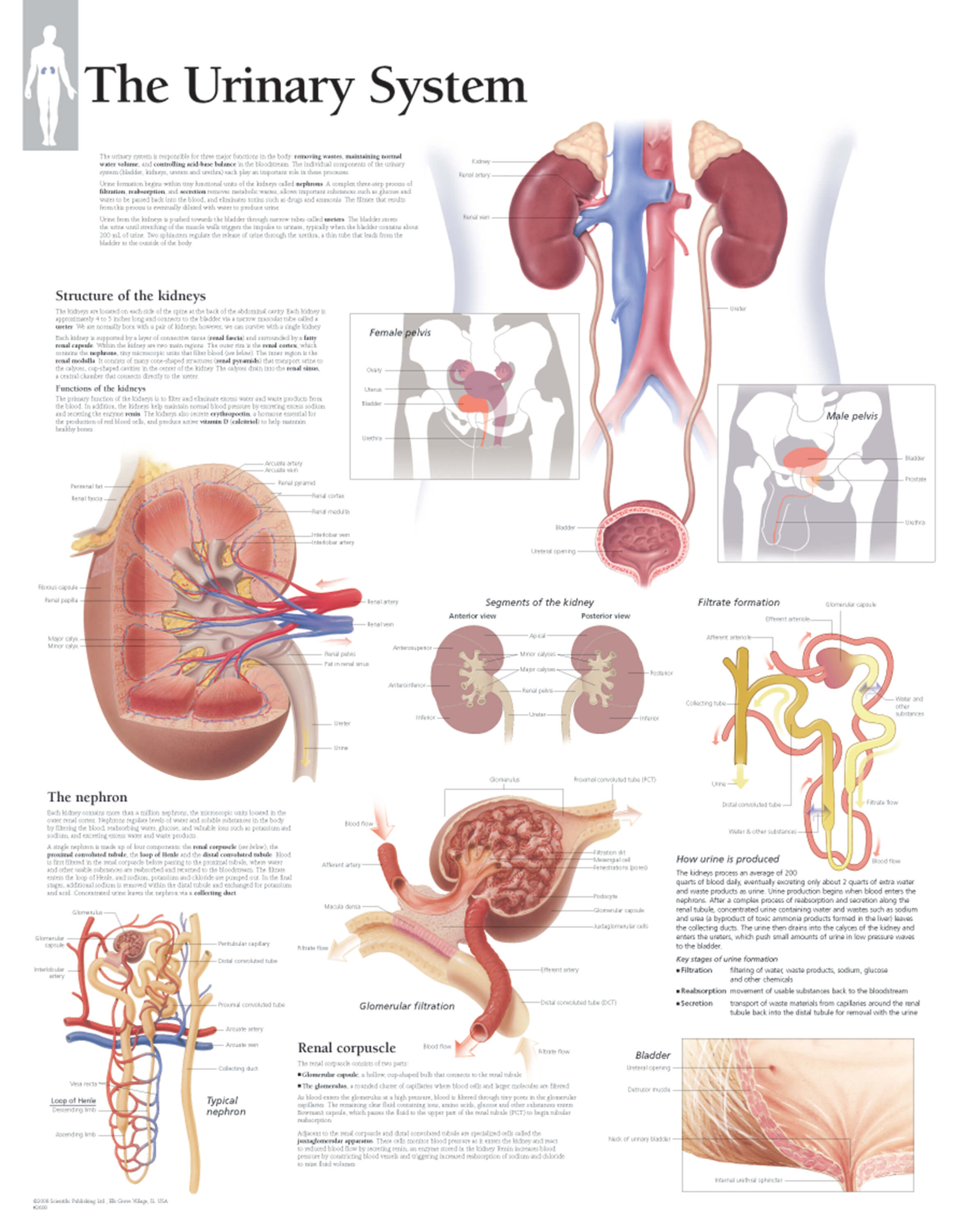
Diagram Of Urinary System
![]()
Urinary system Organs, anatomy and clinical notes Kenhub

Illustration of Urinary System Stock Image F031/5298 Science
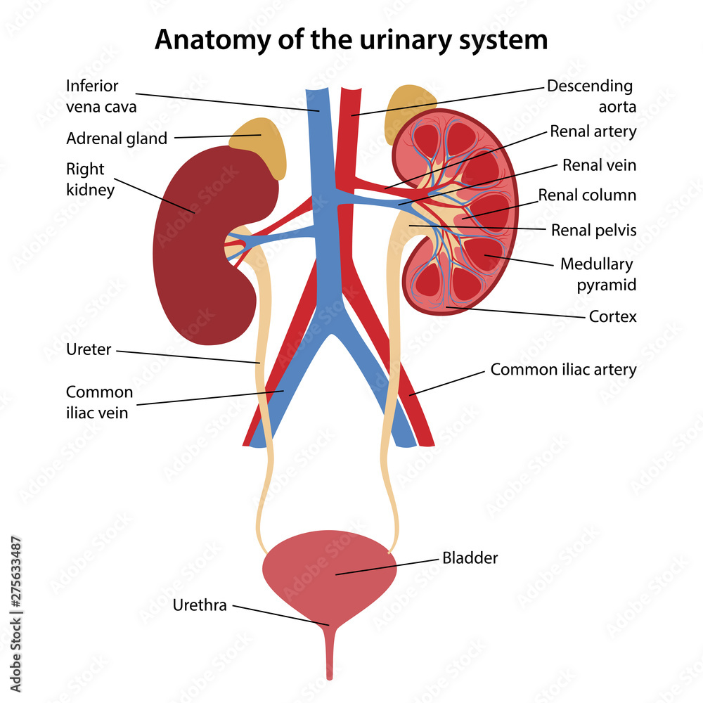
Anatomy of the human urinary system with main parts labeled. Vector
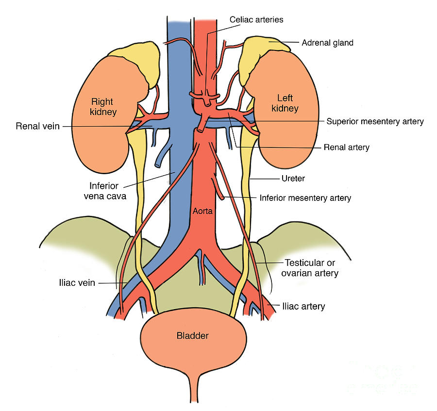
Illustration Of Urinary System Photograph by Science Source
Web Study With Quizlet And Memorize Flashcards Containing Terms Like Renal Artery, Renal Hilum, Kindey And More.
Supplied By Renal Artery And Vein.
Web In This Laboratory, You Will Use Models, Diagrams And Histological Samples To Study The Anatomy Of The Urinary System.
(Portal Systems Also Link The Hypothalamus To The Anterior Pituitary, And The Blood Vessels Of The Digestive Viscera To The Liver.)
Related Post: